Tuesday, 27 February 2018

ACCOMMODATION of EYE :- Mechanism


The Accommodation reflex is the reflex action of the Eye.
Our Eyes have a unique mechanism by which we can even  focus the diverging rays coming from a near object on the retina in a bid to see clearly, mechanism called Accommodation.
the accommodation is the ability to increase the refractive power of optical system of eye by changing the shape of lens.

FAR POINT - The farthest point from the eye at which images are clear (at rest of accommodation).

                                    showing far point in emmetropic eye(A) , hypermetropic eye(B),                                                                   and myopic eye(C)

NEAR POINT- The nearest point at which small objects cab be seen clearly .

RANGE OF ACCOMMODATION- The distance between the near point and far point .

AMPLITUDE OF ACCOMMODATION-  The difference between the dioptric power needed to focus at near point (P) and to focus at far point (R).

*Far and Near point of the eye vary with the Static refraction of the eye.
*In hypermetropic eye far point is virtual lies behind the eye.
*In myopic eye it is real and lies in the front of the eye.
*In emmetropic eye , far point is Infinity and near point varies with age , 7 cm at age of 10 yrs, 25 cm at age of 40 yrs, 33 cm at age of 45 yrs.

Level of Amplitude of Accommodation at different ages-
  • Adolescents have 12-16 D of accommodation.
  • Adults have 4 D at the age of 40 yrs.
  • After 50 yrs of age accommodation reduces at 2 D.
DEPTH OF FIELD - When an object is accurately focused monocularly , the object somewhat near and far are also seen clearly without any change of accommodation, range of distance from the eye in which an objet appears clear known as Depth of field.

DEPTH OF FOCUS - The range at the retina in which an optical image may move without impairment of clarity is known as Depth of focus.


MECHANISM OF ACCOMMODATION :-

The process of accommodation is achieved by a change in the shape of the lens.

THE RELAXATION THEORY (Helmholtz theory)- The relaxation theory also known as capsular theory , most widely accepted .
  • When the eye is at rest (unaccommodated) the elastic substance of young lens is compressed in its capsule by the tension of the zonules. The surfaces of the compressed lens are curved and it changes the dioptric power of the crystalline lens.
  • When zonules are kept under tension by a pull executed on them by the elastic choroid.
  • Contraction of the ciliary muscle causes the ciliary ring to shorten and move forward the equator of the lens. as a result, the zonules are relaxed, the tension on the capsule is relieved and the lens attains a more spherical shape.


OCULAR  CHANGES  IN  ACCOMMODATION :-

1- Changes in Zonules - They slacken during accommodation due to contraction of ciliary muscle.


changes in the ciliary body ring, zonules and shape of lens during accommodation.


2- Changes in Lens -
  • Changes in the curvature of lens.
  • Anterior Pole of the lens moves forward carrying the iris with it.
  • Axial thickness of the lens is increased owing to forward movement of the Anterior pole.
  • Changes in the tension of lens capsule
  • Lens sinks down .
3- Other changes-
  • Pupillary constriction and convergence of eyes.
  • Choroid is stretched forward by the ciliary muscle contraction.
  • Ora serrata moves forward 0.05 mm with each diopter of accommodation.

STIMULUS FOR ACCOMMODATION - Important stimulus are as follows-
  • Apparent size and distance of object.
  • Image blur.
  • Chromatic Abberrations.
  • Scanning movement of the eye.
  • Oscillation of Accommodation.
REACTION TIME:- It is refers to the time lapse between the presentation of an accommodative stimulus and occurrence of the accommodative response.
  • Average reaction time for 'far-to-near' accommodation is 0.64 sec.
  • Average reaction time for 'near-to-far' accommodation is 0.56 sec.
  • Reaction time of convergence response is about 0.20 sec.


LENS :- Definition, Function


*lens is transparent, biconvex, crystalline structure .
*It is placed between iris and vitreous humour.
*The lens capsule is a small cavity or potential space are called as Retrolental.
*Equatorial diameter of the lens at birth about 6.5 mm, increase to 9-10 mm in the second decade.
*Thickness varies with age between 3.5 mm at birth and 5 mm at extreme of age.
*Weight of the lens is about 65 mg at birth.
*Lens has two surface : the anterior surface less convex than the posterior
*The anterior pole is about 3 mm from the back of the cornea.
*Refractive index of the lens is 1.39
*Refractive power of the lens is 16-17 D.
*Accommodative power varies with age , 14-16 D at birth ; 7-8 at 25 yrs of age and 1-2 D at 50 yrs of age.
*Color of the lens is also change with age , transparent lens in Infants and Young adults is colorless.


STRUCTURE OF THE LENS :-


*it consist of three distinct parts-
  1. Lens capsule
  2. Anterior lens epithelium , and
  3. Lens fibres
1- Lens Capsule-
  • it is the component of the globe of the eye
  • it is thin, transparent, hyaline collagenous membrane.
  • the lens capsule is highly elastic.
  • it does not contain any elastic tissue.
  • it is secreted by the basal cell area of the lens epithelium
  • lens is composed of type IV collagen and 10% glycosaminoglycans.
  • it contains enzyme, ATP and glycolytic intermediates.
  • capsule thickness varies according to the age.


2- Anterior Lens Epithelium-
  • It is a single layer of cuboidal nucleated epithelial cells.
  • ALE lies deep to the anterior capsule extending upto equatorial lens bow.
  • it contain three cytoskeletal elements, microfilament, intermediate filaments, and microtubules.
  • the anterior lens epithelium can be divided into three zones : 
  • Central Zone - it consist of cuboidal cells are polygonal in flat section. these cells approx. 10 µm high and 15 µm wide . their nuclei are round and located slightly apically.
  • Intermediate Zone - it consist of comparatively smaller and more cylindrical cells located peripheral to the central zone. their nuclei are round and central.
  • Germinative Zone- it consist of columnar cells which are most peripheral and located just pre-equatorial. Nuclei of these cells are flattened and lie in the plane of cell axis.
3- Lens Fibres-
  • lens fibres from the bulk of the lens.
  • they are long, thin, transparent, firmly packed.
  • Diameters between 4-7 mm and length upto 12 mm.
  • fibres are almost hexagonal in shape and are bound together by the ground substance.
  • the cytoplasm of the cells of the superficial bow region and the newly formed lens fibres contain a Nucleus, polysomes, mitochondria, ER, Golgi apparatus.
  • The nuclei of the lens fibres are present temporarily and disappear later on.

  • the initial fibres forming the fetal nucleus just surrounding the embryonic nucleus are arranged in such a way that they terminate with two Y-shaped sutures on the anterior and the posterior surface of the lens. 


  • Lens fibres are arranged compactly as nucleus and cortex of the lens.
       A- Nucleus- it is the central part containing the oldest fibres.
             it consist of different zones-
  • Embryonic nucleus- is its innermost (formed at 1 to 3 months of gestation).
  • Fetal nucleus- corresponding to lens from 3 months of gestation till birth.
  • Infantile nucleus- corresponding to lens from birth to puberty.
  • Adult nucleus- corresponding to the lens in adult life.
       B- Cortex- it is the peripheral part of the lens substance which lies just outside the adult nucleus.it comprises the youngest (most recent formed) lens fibres.

FUNCTIONS :-
  1. It helps in Accommodation.
  2. it helps in refraction.
  3. it provide transparency.
  4. Nourishment.

Monday, 26 February 2018

AQUEOUS HUMOUR :- Definition , Outflow system, Function


*The aqueous humour is a transparent , watery fluid similar to plasma.
*it containing low protein concentration.
*it is secreted from the ciliary epithelium.
*It is involved with all portion of the eye.
*the principal ocular structures concerned with it are:
  1. ciliary body
  2. posterior chamber
  3. anterior chamber
  4. angle of the anterior chamber, and
  5. aqueous outflow system.


CILIARY BODY:-

*it is the site of the aqueous production.

POSTERIOR CHAMBER:-

*it contain about 0.06 ml of aqueous humour .
*the fresh formed aqueous humour from the ciliary process collected in this space.

Compartments of posterior chamber.

*it divided in to three compartments-

1- Prezonular space. it lies between the posterior surface of the iris and anterior surface of the zonular fibres.

2- Zonular space. it is bounded centrally by equator of the lens, peripherally by the ciliary processes, anteriorly by the posterior surface of anterior zonular fibres and posteriorly by anterior surface of posterior zonular fibres.

3 - Retrozonular space. it lies between the posterior surface of zonules and the peripheral part of anterior vitreous face.


ANTERIOR CHAMBER :-

*it is about 3 mm deep in the center of adults.
*it is comparatively shallow in very young children and old people.
*it contains about 0.25 ml of the aqueous humour.
*through pupil it communicates with posterior chamber.

ANGLE OF THE ANTERIOR CHAMBER:-

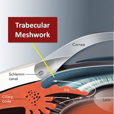
*it is the angle recess formed in between posterior surface of cornea and anterior surface of iris bounded from anterior to posteriorly by-
  1. Schwalbe's line
  2. Trabecular meshwork
  3. Sclera spur
  4. ciliary band
1- Schwalbe's line -
  • it is formed by the prominent end of the descemet's membrane of the cornea
  • it is a fine ridge seen just in front of the trabecular meshwork.
  • width 50-150 µm.
2- Trabecular meshwork-
  • it is seen as a band just anterior to the sclera spur.
  • it has no pigment at birth .
  • pigmentation acquired with age.


3- Scleral spur-
  • it is the posterior portion of the scleral sulcus.
  • it is wedge shaped circular ridge.
  • it lies posterior to trabecular meshwork


4- Ciliary  band-
  • it is the most posterior part in the angel recess.
  • width depend on the level of iris insertion
  • wide in myopes and narrow in hypermetropes
  • it appears as a grey/dark brown band.

AQUEOUS OUTFLOW SYSTEM:-


it includes the -
  1. Trabecular meshwork
  2. Schlemm's canal
  3. Collector channels
  4. Aqueous veins
  5. Episcleral veins

1- Trabecular meshwork-
  • it is a sieve like structure.
  • aqueous leave the eye from here.
  • it bridges the scleral sulcus and converts in into tube.
2- Schlemm's canal-
  • it is an endothelial lined oval channel present.
  • it is single channel but occasionally branches into a plexus-like system.
3- Collector channels-
  • it is lined by vascular endothelium .
  • they are relatively wide at their origin 60-90mm.
  • no valves are present in the collector channels.
  • also known as intrascleral aqueous vessels
  • these intascleral aqueous vessels can be divided into two system-
  • 1- Direct system
  • 2- Indirect system


4- Episcleral veins -
  • most of the aqueous vessels drain in to episcleral vein.

FUNCTIONS:-
  1. Maintenance of intraocular pressure.
  2. Metabolic rate
  3. Optical function
  4. Clearing function


CHOROID :- Definition, Function, Microscopic structure


Choroid is the vascular layer of the Eyeball.
It is the Posterior most part of the vascular layer of the eyeball.
Its inner surface is smooth, brown and lies in contact with pigment epithelium of the Retina.
It extends from the optic disc to ora serrata.
The outer surface is rough and lies in contact with sclera .
It is thicker posteriorly - 0.22 mm
Thicker at Anterior - 0.1 mm.


MICROSCOPIC STRUCTURE :-

It consists the following four layer -

*Suprachoroidal lamina (lamina fusca)
*Stroma of the choroid.
*Choriocapillaris.
*Basal lamina (Bruch's membrane or lamina vitrae).

1- Suprachoroidal lamina -

It is thin membrane 10-34 mm.
It consists condensed collagen fibres, fibroblasts and melanocyte.
Transition zone of choroid and sclera.
Blends with choroid and lamina fusca of sclera.

2- Stroma of the choroid -

It contains plenty of pigment cells .
It contains other cells like microphage, masts cells, plasma cells and lymphocytes.
It consists of loose collagenous tissue .
Arteries are branch of short posterior Ciliary arteries.

3- Choriocapillaris -

It consists of a rich capillary Network.
It nourishes the pigment epithelium and the outer layers of sensory retina.
Capillaries are fenestrated.
Extends anteriorly to ora serrata.
Greatest density of capillaries at macula.

4- Basal lamina (Bruch's membrane or lamina vitrae) -

It is inner most layer of choroid and approx- 2-4 mm in thickness.
It is multilayered structure.
It lies between the Choriocapillaris and pigment epithelium of the Retina.
It prevents choroid vessels from penetrating the Retina but allow nutrients, protein etc.

It is described to consists of five layers :

*Basement membrane of the retinal pigment epithelium.
*An inner collagen layer.
*A middle elastic layer.
*An outer collagen layer.
*The basement membrane of the Choriocapillaris.


FUNCTION:-

  1. it gives protection to the eyeball.
  2. supply of nutrients to tissues of the eyeball.
  3. converging the light.

Sunday, 25 February 2018

CILIARY BODY :- Definition, microscopic structure


*The Ciliary body is the part of the Uvea, The layer of tissue that delivers oxygen and nutrients to the eye tissues.
*Ciliary body is the forward continuation of the choroid at ora serrata.
*The inner side of the triangle is divided into two parts -

*Pars plicata - the Anterior part having finger like process .
*Pars plana - the Posterior smooth part (about 5 mm wide temporally and 3 mm wide at nasally).

MICROSCOPIC STRUCTURE :-

It consists of following five layers -

*Supraciliary lamina.
*Stroma of the ciliary body.
*Layer of pigmented epithelium.
*Layer of non-pigmented epithelium.
*Internal limiting membrane.


1- Supraciliary Lamina -

*Outermost condensed part of the stroma .
*It consists of collagen fibres.
*It is a continuation of the suprachoroidal lamina ,posteriorly.
*Anteriorly it continues with anterior limiting membrane of the Iris.

2- Stroma of the ciliary body -

*It consists of connective tissue of collagen and fibroblasts.

3- Layer of pigmented epithelium -

*It is forward continuation of the RPE (Retina pigmented epithelium).
*Anteriorly it is become continuation with the Anterior pigmented epithelium of the Iris .

4- Layer if non-pigmented epithelium -

*It consists of cuboidal cells and low columnar cells.
*Forward continuation of the sensory retina which stop at the ora serrata .

5- Internal limiting membrane -

*It is the forward continuation of the internal limiting membrane of the Retina.


CILIARY PROCESSES :-


*Folds on the inner Ciliary epithelium are called ciliary processes.
*Ciliary processes are arranged in a circle, and form a sort of frill behind the Iris .
*It is composed of three basic components :
           *The network of capillaries.
           *Stroma of ciliary processes.
           *Two layers of epithelium.

 FUNCTION:-
  1. It helps in production of aqueous humour.

Saturday, 24 February 2018

IRIS :- Definition , function, Microscopic structure.


*Iris is the Anterior most part of the UVEAL TRACT.
*The average diameter and thickness of Iris is 12 mm and 0.5 mm.
*In the center of Iris "slightly Nasal" , is a hole or aperture called PUPIL (3-4 mm).
*Pupil control the amount of light reach the Retina.
*Iris lies between the Cornea and lens into Anterior to posterior chamber.
*Iris is attached to the middle of anterior surface of the ciliary body at periphery.
*Iris is thinnest at its root( tears away easily from ciliary body).


MICROSCOPIC APPEARANCE :-


Anterior part of the Iris -

*IRIS in Anterior divided into a Ciliary Zone and a Pupillary zone by zigzag line called COLLARETTE (thickest region of the Iris).

1- CILIARY ZONE -

* Radial streaks are straight when pupil is small and wavy when it is dilated.
*Crypts are depression where the superficial layer of Iris is missing.
crypts are arranged in two rows -

A- Peripheral crypts
B- Central crypts.

*Contraction furrow are the tenuous line concentric to the collarette.

2- PUPILLARY ZONE- 

*This zone lies between the collarette and pigmented pupillary frill.
*It is about 1.6 mm wide.

*Pigment frill is a fringe of black pigment present at the pupillary margin.

Posterior part of the Iris -

*Posterior part of the Iris is dark brown or black in color.
*It looks smooth with naked eye.
*Following radial , circular furrows and folds are present in posterior surface of Iris.

MICROSCOPIC STRUCTURE :-


Anterior to posterior it consists four layers  are as follows -
*Anterior limiting layer
*Iris Stroma
*Anterior epithelial layer
*Posterior pigmented epithelial layer.

1- Anterior limiting layer -

*The definitive color of the Iris depends upon this layer.
*Blue Iris - layer is thin and contain few Pigment cells.
*Brown Iris - thick layer and doubly pigmented.


*It is the Anterior most condensed part of the Stroma.
*It consists of melanocyte, and fibroblasts.

2- Iris Stroma -

*It form the main bulk of the Iris tissue.
It consists of loosely arranged collagenous Network.


3- Anterior epithelial layer -

*It is the Anterior continuation of the pigment epithelium of retina and ciliary body.
*Lacking of melanocyte.

4- Posterior pigmented epithelial layer -

*This layer is derived from the internal layer of the optic cup.
*It is the continuation of the non- pigmented epithelium of the Ciliary body.

FUNCTION :-
  1. It is responsible for controlling the diameter and size of the pupil.
  2. Eye color is defined by that of the iris.
  3. Maintains the gradient of Intraocular pressure between the front and back of the eye


Thursday, 22 February 2018

LIMBUS:- Definition


The corneal Limbus is the border of the cornea and sclera.
It refers to a circumcorneal transitional zone of the conjunctivocorneal and corneoscleral junction.
It associated with two junction -

1- Conjunctivocorneal junction.
2- Corneoscleral junction.

*CONJUNCTIVOCORNEAL JUNCTION :-

°At this point, the bulbar conjunctiva is a tightly adherent to the underlying structures.
°The Substantia propria of the Conjunctiva stops here but it is epithelium continues with that of the cornea.
°The cells contains melanin and differ from those of the cornea in having no prickles between them.
°Limbal epithelium is similar to corneal epithelium but its basal cell are arranged in peculiar pattern called as pallisade of vogt, containing stem cells (these stem cells are unipotent stem cells).


*CORNEOSCLERAL :-

°It is the transition region between the Cornea and sclera.
°It is highly vascular.
°It is 1.5 mm width.
°It contains -
 * Trabecular meshwork
 *Canal of schlemm

°At this junction, the transparent corneal lamellae become continues with the oblique , circular and opaque fibres of the sclera.

SURGICAL LIMBUS :-

It is a 2 mm wide circumcorneal transitional zone between cleat cornea on one side and the opaque  sclera on the other side.
  
Border of surgical Limbus :-

1- Anterior limbal border -

° it is the antr boundary of the surgical limbal seen form the exterior.
°Marked by Prominent ridge created by Insertion of the Conjunctiva and Tenon's capsule.
°The antr limbal border overlies the termination of the Bowman's membrane.

2- Mid limbal line -

°Junction of the blue zone with white area.
Overlies the termination of the Descemet's membrane.
°Most useful external landmark.

3- Posterior limbal border -

°It lies about 1 mm posterior to the mid Limbal line .
°It overlies the scleral spur and can only be seen with use of sclerotic scatter illumination.
°It forms the posterior border of the surgical Limbus.

Zones of surgical limbus :-

1- Blue Limbal zone -

°It is bluish translucent zone seen posterior to the antr limbal border.
°Adjacent to this bluish zone posterior is the white sclera.
°The extent of blue Limbal zone is about 1mm in superior quad , 0.8 mm in inferior quad and about 0.4 mm in nasal and temporal quad.

2- White Limbal zone -

°It is a 1 mm wide whitish area which lies between the mid-limbal line and the posterior limbal border.
°Overlies the the trabecular meshwork .
°The total width of the surgical limbus is greatest in the superior quad where width of blue zone is Max.


Tuesday, 20 February 2018

CORNEA :- Definition, Function, Microscopic structure

*Cornea is the one the most important part of the eye ,it allow to enter the light in the eye.
*It is a transparent, avascular, watchglass like structure.
*It forms anterior 1/6th part of the fibreous layer of the eyeball.

DIMENSIONS -

Horizontal diameter(Anterior) :-  11.75 mm
Vertical diameter(anterior) :-  11 mm
Thickness (center )  :-  0.52 mm
Thickness (periphery)  :-  0.67 mm
Anterior Radius of curvature  :-  7.8 mm
Posterior radius of curvature  :-  6.5 mm
Refractive power (anterior) :-  +48 D
Refractive Power (posterior)  :-  -5 D
Net refractive power of cornea  :-  +43 D
Refractive index  :-  1.37

HISTOLOGY :-

The cornea was considered to consists of five distinct layer, from anterior to posterior are :-

* Epithelium
* Bowman's membrane
* Stroma
* Descemet's membrane
* Endothelium
* Pre-descemet's membrane (dua's layer) {recently found , it lies between Stroma and descemet's membrane}


EPITHELIUM :-

*It is about 50-90 Âµm thick (10% of corneal thickness)
*It consists of 5-7 layers of cell.
*It is composed by Stratified squamous non-stop keratinized.
*It becomes continue with epithelium of Bulbar Conjunctiva at the Limbus.

BOWMAN'S MEMBRANE :-

*It is about 8-14 Âµm in thickness .
*It attach to the corneal Stroma.
*It is consists of Acellular mass of condensed collagen fibres( type I and IV)
*It acts as a smooth base for epithelium uniformity , helps in refraction.

STROMA ( substantia propia) :-

*It consists of collagen fibrils .
*Cell embedded in hydrated matrix of proteoglycans.
*It is about 0.5 mm in thickness (90% of total thicknesses of cornea).

DESCEMET'S MEMBRANE :-

*It represents the Basement membrane of the corneal epithelium.
*It composed by Collagen and glycoproteins ( no elastic fibres)
*It's thickness varies with age , 3 Âµm at birth and 10-12 Âµm in young adult.
*It is very resistant to Chemical agent , Trauma , InfevInfe, and Pathological processes.
*It is a strong Homogeneous layer.
*It is separated from the Stroma by pre- descemet's membrane.

Endothelium :-

*Endothelial cells are attached to the descemet's membrane.
*It contains an active pump mechanism.
*Involved in active secretion and protein synthesis.
*It consists of a single layer of Flat polygonal cells .

BLOOD SUPPLY OF THE CORNEA -

*The cornea is an avascular structure.
*Small loops derived from the Anterior ciliary vessels invade it's periphery for about 1 mm and provide nourishment.( loops are not in the cornea but in the subconjunctival tissue which overlap the Cornea.)

NERVE SUPPLY OF THE CORNEA -

*The cornea has rich supply of sensory nerve endings derived mainly from the long ciliary nerves which are branches of the nasociliary nerve.

FUNCTION:-
  1. It provide protection to eye from foreign bodies. 
  2. It helps to refract the light.
  3. it focuses the entry of light into the eye.

ACCOMMODATION of EYE :- Mechanism

The Accommodation reflex is the reflex action of the Eye. Our Eyes have a unique mechanism by which we can even  focus the diverging...